
Introduction: A case study is presented which illustrates how gout can be overlooked as a cause of posterior ankle pain in primary care settings.
Case presentation: A case of gout affecting the posterior ankle is presented. The case discussed was referred to a physiotherapy service as suspected bursitis and tendonitis, following consultant referral and subsequent MRI, but after minimal improvements were seen, further assessment revealed the possibility of gout as the cause of symptoms.
Conclusion: This case study discusses an alternative diagnosis for posterior ankle pain that is not responding to physiotherapeutic management.
Introduction
Gout is one of the best understood systemic, rheumatic diseases of the 21st century1. Having been extensively studied in the medical community, gout is generally well managed once a diagnosis has been made and confirmed. However, despite the extensive literature gout remains one of the most frequently mismanaged and misdiagnosed diseases in both emergency departments and primary care settings 2,3.
The following case report outlines the case of a 58-year old patient referred to physiotherapy with acute, posterior ankle pain, possibly bursitis or tendonitis from a consultant orthopaedic surgeon.
The report highlights the importance of obtaining a detailed history, understanding the clinical presentation of gout and how it reacts to physiotherapeutic management so that it is detected early and managed appropriately. More importantly, it highlights the importance of not relying on MRI scans in isolation and ensuring each clinician make a full examination regardless of scans and where the referral came from.
Epidemiology
Epidemiology studies show that prevalence of gout has risen in the last 10 years 4. This may be attributable to increased longevity, improvements in medical care and changes in lifestyle 5.
Risk factors associated with gout include obesity, alcohol consumption, high purine content in diets, hypertension and use of certain predisposing medications such as aspirin and thiazide diuretics 1. Gout is most commonly seen
in individuals over the age of 40, prevalence increases with age 6 and men are at a higher risk of gout than women 7.
Pathology
Gout is an inflammatory disorder characterised by the deposition of monosodium urate crystals, an end product of purine metabolism. Crystal depositions can occur in the joints and in soft tissues, leading to an inflammatory response 8.
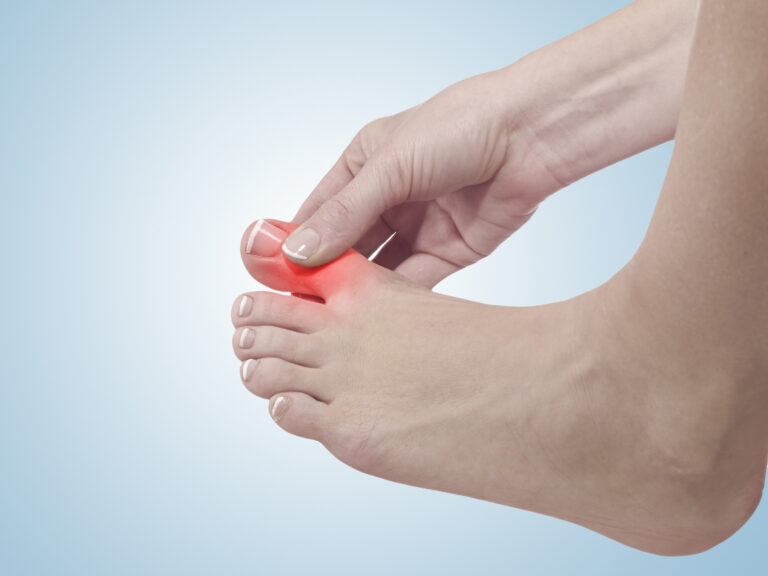
Patients with gout typically present with rapid onset of severe pain, accompanied by swelling and erythema. Joints in the lower limbs are more commonly affected 9, in particular the metatarso-phalangeal joint of the great toe, which is the first joint affected in more than 50% of patients. Differential diagnosis of acute gout may also include tenosynovitis, tendonopathy, bursitis or cellulitis 10.
Acute flare-ups of gout commonly resolve in 10 days if diagnosed early and treated correctly 11, 12.
Between episodes of gout, patients generally enter an asymptomatic stage but if left untreated, episodes often become more frequent and of longer duration 9. The more advanced stage of the disease is characterised by chronic destructive arthritis, secondary degenerative changes and tophi 13. Untreated cases can also lead to reduced foot function and abnormal gait characteristics secondary to pain avoidance strategies 14.
Management
For acute episodes of gout, correct diagnosis is paramount for successful management. Identifying risk factors, in particular considering the patient’s age and if they have a history of gout, or have any family history, is important.
Initial treatment should include rest and prompt medication consisting of full doses of a non-steroidal anti-inflammatory drug (NSAID). It is recommended that aspirin be avoided as it causes retention of uric acid in lower dosage. For more chronic cases of gout, preventative medical management consists of prolonged administration of drugs to lower serum urate levels 10.
Case Presentation
A 58-year-old man was referred to our physiotherapy service, by a consultant orthopaedic surgeon, with a 3-week history of left sided, posterior ankle pain. The initial diagnosis from the consultant was Achilles tendonitis/Bursitis.
Pain was initially very localised and acute but had since settled to a generalised discomfort around the back of the left ankle. The pain was aggravated by gait and pressure on the left heel during heel strike and toe off.
Subjective assessment revealed no history of trauma but the patient reported an area of swelling on the back of his heel, which had been present for some years. No noticeable change in the size of the swelling occurred over recent months but this was the focal point of the pain.
Examination of the heel confirmed the presence of a swelling over the calcaneal tuberosity, which was bony hard and tender on palpation. No other abnormalities were found in the Achilles tendon (TA) itself, nor did examination of the joints around the ankle reveal any significant findings. All resisted movements were normal, with some slight pain on resisted plantiflexion of the foot, worse in weight bearing.
An MRI scan revealed a large calcaneal spur and some soft tissue swelling around the adjacent bursa but neither MRI nor Xray showed evidence of bony swellings. No blood tests were done.
The initial findings suggested that the symptoms where caused by Achilles tendonitis and inflammation of an adventitious bursa between the TA and skin. As the attachment site of the TA, the calcaneal tuberosity is considered to be among the skeletal parts of the human body most exposed to wear and tear 15 and so irritation of the bursa and surrounding soft tissues is not uncommon. Initial treatment given consisted of gentle massage, ultrasound, interferential therapy and acupuncture to reduce pain and stimulate healing followed by strapping and a heel pad to relieve irritation of the bursa.
The patient had been taking over the counter anti inflammatories (Ibuprofen 200mg three times daily)since the start of symptoms but these were proving ineffective at reducing symptoms. The patient was on no other medication at the time of assessment.
A review of the patient after several treatment sessions revealed minimal improvement overall and the patient reported symptoms of increased pain over the posterior heel spur following treatment. It was also noted that the spur was warm to touch and very red which was not present on the initial examination.
As massage and acupuncture were reported to have aggravated symptoms, they where immediately stopped. Further subjective assessment revealed the patient had a family history of gout. In consideration of this and given his age, gender and presentation, further investigation into gout as the cause of the complaint was recommended. Treatment was modified to only interferential therapy for pain relief and advice given to limit activity involving excessive weight bearing on the left foot until the following session.
Discussion
It is important get an accurate diagnosis for posterior ankle pain prior to commencing therapy to avoid unnecessary aggravation of symptoms, as this case study has demonstrated. Identifying gout as the cause of symptoms is a vital step towards establishing a lifelong treatment plan, which may include medication to prevent future episodes.
The presence of monosodium urate crystals is general considered to be gold standard for the diagnosis of gout but this is not often a procedure available to Physiotherapists. Therefore, physiotherapists must rely on subjective questioning to identify gout as a possible cause of symptoms, which can lead to a recommendation for investigation to confirm it or rule it out. Gout is commonly seen in the 1st MTP joint, but can also affect other weight bearing joints such as the Heel, Calcaneal bursa and knee 3.
A number of proposed clinical criteria for the diagnosis of gout have been put forward in the literature but with little evidence to support their use as a definitive diagnostic tool. A review of six proposed clinical criteria showed that they had a sensitivity of 87.6% but 19.5% of other diseases would be misdiagnosed as gout 18, 19. It is therefore questionable whether gout can be differentiated from other pathologies using clinical criteria alone but does suggest that understanding clinical signs and symptoms reduce the chance of this condition being missed in primary care settings.
Conclusion
This case report discusses the clinical presentation of gout and how its clinical presentation may lead clinicians to believe that symptoms are from other, more commonly seen pathologies leading to posterior ankle pain. It has particular relevance to Physiotherapists and other practitioners acting as a first point of contact for musculoskeletal disorders and is believed to be the first case report describing gout as a cause of posterior ankle pain in the literature.
References
1. Bieber JD, Terkeltaub RA. Gout: on the brink of novel therapeutic options for an ancient disease. Arthritis Rheum 2004;50:2400–14.
2. Chin MH, Wang LC, Jin L, et al. Appropriateness of medication selection for older persons in an urban academic emergency department. Academy of Emergency Medicine 1999;6(12):1189-1193.
3. Chen L, Schumacher R. Gout: can we create an evidence-based systematic approach to diagnosis and management? Best Practice & Research Clinical Rheumatology 2006;20(4):673-684.
4. Richette P, Bardin T. Gout. Lancet 2010;375:318-29.
5. Saag KG, Choi H. Epidemiology, risk factors, and lifestyle modifications for gout. Arthritis Research and Therapy 2006;8(1):1181-1907.
6. Van Doornum S, Ryan PF. Clinical manifestations of gout and their management. Med J Aust 2000;172(10):493-497.
7. Agudelo CA, Wise CM. Crystal-associated arthritis in the elderly Rheum Dis Chin North Am 2000;26(3):527-546.
8. Merriman TR, Dalbeth N. The genetic basis of hyperuricaemia and gout. Joint Bone Spine 2011;78(1):35-40.
9. Kim KY, Schumacher HR, Hunsche E, Wertheimer AI, Kong SX. A literature review of the epidemiology and treatment of acute gout. Clin Ther 2003; 25:1593–617.
10. Nuki G. Gout Review Article. Medicine 2006; 34 (10): 417-423. http://www.sciencedirect.com/science?_ob=MImg&_imagekey=B82YB-4M04F8G-1-9&_cdi=33054&_user=9164652&_pii=S135730390600048X&_origin=search&_zone=rslt_list_item&_coverDate=10%2F31%2F2006&_sk=999659989&wchp=dGLbVlz-zSkzk&md5=cf39166c6998d743af389027841f6ae3&ie=/sdarticle.pdf (Accessed 520/05/11).
11. Braunwald E, Fauci AS, Kasper DL. Harrison’s Principles of Internal Medicine (15th ed.). New York: McGraw-Hill 2001: 649.
12. Cheng TT, Lai HM, Chiu CK. A single-blind, randomized, controlled trial to assess the efficacy and tolerability of rofecoxib, diclofenac sodium, andmeloxicam in patients with acute gouty arthritis. Clinical Therapy 2004;26(3):399-406.
13. Teng G, Nair R, Saag K. Pathophysiology, Clinical Presentation and Treatment of Gout. Drugs 2006;66(12):1547-1563.
14. Rome K, Survepalli D, Sanders A, Lobo M, McQueen F, McNair P, Dalbeth N. Functional and biomechanical characteristics of foot disease in chronic gout: A case-control study. Clinical Biomechanics 2011;26(1):90-94.
15. Kachlik D, Baca V, Cepelik M, Hajek P, Mandys V, Musil V. Clinical anatomy of the calcaneal tuberosity. Annals of Anatomy 2008;190:284-291.
16. McGill NW. Gout and other crystal-associated arthropathies. Baillieres Best Pract Res Clin Rheumatol 2000;14:445–460.
17. Zhang W, Doherty M, Bardin T, et al. EULAR evidence based recommendations for gout. Part II: Management. Report of a task force of the EULAR Standing Committee for International Clinical Studies Including Therapeutics (ESCISIT). Ann Rheum Dis 2006;65(10):1312-24.
18. Schumacher HR, Edwards LN, Perez-Ruiz L, et al. Special interest group on outcome measures for acute and chronic gout. The Journal of Rheumatology 2005;32:2452-2455.
19. Schumacher HR, Wortmann RL. Monosodium urate crystal deposition arthropathy. Part I: Review of the stages and diagnosis of gout. Advanced Studies in Medicine 2005;5(3):133-138.